Femur: Anatomy, Parts, Functions And Attachment Muscles
Femur: Anatomy, Parts, Function and Attachment Muscles – What is the function of the femur in humans?, On this occasion About Knowledge.co.id will discuss it and of course other things that also surround it. Let's take a look at the discussion in the article below to better understand it.
Table of contents
-
Femur: Anatomy, Parts, Function and Attachment Muscles
- Anatomy of the femur
- Thighbone Section
- Function of the femur
- Femur Attachment Muscles
- Share this:
- Related posts:
Femur: Anatomy, Parts, Function and Attachment Muscles
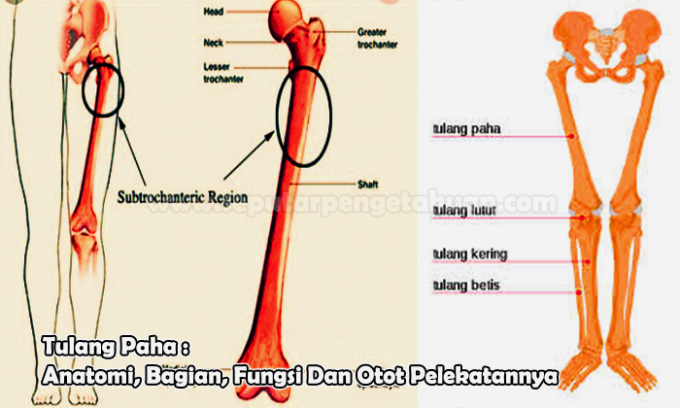
The thigh bone or femur is the largest body part and also the strongest bone in the human body. The function of the thigh bone is to connect the hip and thigh and support the body when walking.
The thigh bone is located in the human thigh between the hip and knee bones. The femur attaches to the hip via a ball and socket joint that moves and articulates the hip.
Anatomy of the femur
The femur is composed of a proximal head and neck and two distal condyles.
The head of the femur will form the joint at the hip. Meanwhile, other proximal parts such as the greater trochanter and the lesser trochanter act as a place of muscle attachment. In the proximal posterior part there is the gluteal tuberosity, which is a rough surface where the gluteus maximus muscle is attached. Nearby is the linea aspera, which is where the biceps femoris muscle attaches.
One of the functions of the head of the femur is as a place for the production of red blood cells (erythrocytes) in the bone marrow.
At the distal end there is a condylar structure along with the knee to form the condylar joint. There are 2 condyles, the medial condyle and the lateral condyle. Both are bounded by a structure called the intercondylar fossa.
Thighbone Section
There are 2 major parts of the femur, the proximal femur and the distal femur.
The location of the proximal part of the femur is near the hip bone (pelvis) and is composed of the head, neck, trochanter, major, minor and median femur. Meanwhile, the location of the distal part of the femur is on the thigh that attaches to the kneecap.
Also Read:Upper Bone Function: Part, Structure and Discussion
Function of the femur
In addition to being known to be large and strong, the femur is also the longest bone in the human body.
Then, what are the uses of the thigh bone that humans really need for this activity?
- Strongest bones
Like the strongest and strongest bone in the human body, the thigh bone is very vital, in supporting the body. The femur also protects the stability of the human body.
Not only that, when humans are carrying heavy loads, the femur also helps so that the body is always strong to support the burden. This is because the femur can hold up to 30 times the weight of the human body.
No wonder the femur is said to be the largest and strongest bone in the human body. Not only that, the thigh bone is also sturdy to withstand the strength of up to 800 kg to 1 ton.
That's why, the femur is not easily broken. Even if the femur is broken, usually only things like a car accident or a fall from a height can cause it. At the very least, it will take around 3-6 months for the femur to recover from the fracture.
- Articulation and leg energy
Its "strategic" position makes the use of the thigh bone very diverse. One of them is to produce articulation and leg power, for running, walking, and standing.
The very top of the femur, which is a ball, connects to the hip joint. So, the legs can move in all directions.
- The main bone in the leg
Not only big and strong, the femur is also the main bone of the foot, which is the support of all the bones in the leg.
Because, the distal (base) of the thigh bone, so the position of the attachment of all the leg bones, from the knee to the foot is very basic.
- Place of manufacture of red blood cells
The medullary cavity, located in the femur, is where red blood cells are stored and made.
In the medullary cavity, there is bone marrow, which has 2 types of stem cells, namely hemopoietic (blood cell producing) and stromal (fat producing).
- Where to attach the knee
Also Read:Electromagnetic Waves: Definition, Properties, Formulas, Benefits, Spectrum
The very base of the thighbone (distal), is where the patella (kneecap) attaches.
At the base of the femur, there is a lateral condyle, which allows the knee to move freely.
- Lower Body Exercise Equipment
The femur also serves to help the leg move in a straight line and bend towards the hip, so it is like a basic human movement apparatus.
- Together with the knee, make up the condylar joint
At the distal end of the femur is the condyle which forms the condylar joint with the knee. There are 2 condyles, the medial condyle and the lateral condyle.
- Hip and knee joint
The femur also serves as a link between the hip bones and the knee.
- Place of attachment of muscles and pigment
The femur is the place for the big muscles to attach. There are 2 types of muscles found in the femur, namely the origin and insertion muscles.
The origin of the muscle is a muscle that has a normal movement or always when a contraction is attempted.
The femur is the origin for several muscles such as the gastrocnemius, vastus lateralis, vastus medialis, and vastus intermedius muscles.

Femur Attachment Muscles
The following are the muscles attached to the femur, including:
The femur is the origin of a number of muscles such as:
- Vastus intermedius muscle
- Vastus medialis muscle
- Vastus lateralis muscle
- Gastrocnemius muscle
The femur is the insertion of a number of muscles such as:
- iliopsoas muscle
- Gluteus maximus muscle
- Gluteus medius muscle
- Tensor fasciae latae muscle
That's the review from About Knowledge.co.id about Femur: Anatomy, Parts, Function and Attachment Muscles,Hopefully it can add to your insight and knowledge. Thank you for visiting and don't forget to read other articles
