
Definition of Endospores, Mechanisms, Processes, Structures and Types
Definition of Endospore
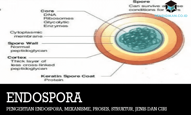
These endospores are resistant structures produced by bacteria to then survive under unfavorable environmental conditions or conditions. These endospores contain DNA as well as a small cytoplasm, which is surrounded by a protective outer covering.
Endospores germinate to produce new organisms when environmental conditions become favorable. Therefore, this endospore is considered a type of reproductive cell. The bacterial genera, Bacillus, Clostridium, and Paenibacillus produce endospores. These endospores can or can survive in harsh conditions such as dehydration, high and low temperatures, chemicals, and UV radiation.
The cell wall of this endospore is composed of dipicolinic acid, which gives the endospore heat-resistant properties. Moist heat treatment at 121 °C for 15 minutes can or may destroy bacterial endospores.
This endospore is an alternative life form produced by Bacillus, Clostridium, as well as some Bacterial genera include Sporosarcina, Sporolactobacillus, Desulfotomaculum, Oscillospira, and Thermoactinomyces. This Bacillus is an obligate aerobe that lives in the soil only while Clostridium is a mandatory species. This anaerobic is often also found as a normal flora of the intestinal tract in an animal. These endospores are then formed by a bacterium, under conditions or environmental conditions that are not beneficial, such as lack of nutrients and water, very hot or very cold temperatures and also poison. This endospore is a thick-walled body and is also very resistant (resistant).
The endospore contains genetic material, a little cytoplasm, and ribosomes. The thick endospore wall is composed of protein and then makes the endospore resistant to light radiation, drought, high temperatures and chemicals. If the environmental conditions are favorable, these endospores will grow into new bacterial cells. These endospores are more resistant to conditions or environmental conditions that are actually less favorable than the vegetative cells of bacteria. The process of spore formation is known as sporulation. If the environmental conditions improve, the endospores will then split into vegetative cells again, which is called the germination process.
The function of the endospore for the bacteria is as a survival structure (dormant structure). These structures that allow bacteria to survive under unfavorable conditions are extreme environments (drought, very low or very high temperatures) or lack of nutrition.
Endospore Characteristics
Most of the bacteria that can or can build endospores are gram-positive bacteria. These gram-positive bacteria are a group of eubacteria whose cell walls absorb violet color during the gram staining process and have or have thick enough peptidoglycan. Bacteria that can or can form endospores, for example, are Bacillus mycoides.
These endospores have or have impermeable properties, then they are able to survive better against bacteria areas of drought, low temperature, high temperature, disinfectant, and also an unfavorable environment other. When the environmental conditions have improved, these endospores will germinate into new vegetative cells.
Types of Endospores
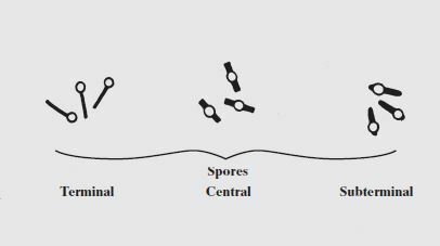
There are 3 types of endospores in bacteria based on their location. The following is a chart, description and examples of bacteria that produce spores of each type.
- Terminal endospore: Endospore which is at one end of the bacterial vegetative cell. Examples are: Clostridium tertium
- Subterminal endospore: Endospore which is positioned at the tip of the cell. However, more towards the center of the cell. An example is: Clostridium perfringens
- Central endospore: Endospore which is in the center of the vegetative cell. Examples are: Clostridium bifermentans
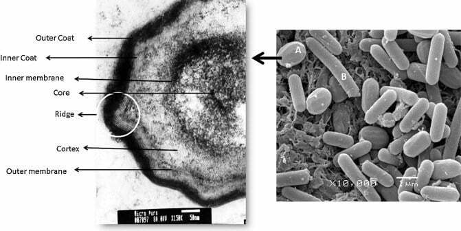
Endospore Structure and Components
When compared with non-endosporous cells (vegetative cells) which appear to have or have a single layer of cell wall, this endospore has more components that build structure endospore. The structure of the endospore includes:
- Exosporium: the outer wall of the endospore which is composed of a thin layer of protein
- Mantle: Several layers of specialized proteins that make up the endospore mantle
- Cortex: Layer composed of peptidoglycan
- Core: The part consisting of the cytoplasm, nuclear wall, ribosomes, circular chromosomes, cytoplasmic membrane, and other vital organelles
The core part of the endospore has or has a gel-like consistency because it contains very little water. This can or may increase the resistance of the molecules inside the endospore from high temperatures (up to 150 °C) and harmful chemicals such as hydrogen peroxide.
Endospore Composing Chemicals
Dipicolinic Acid
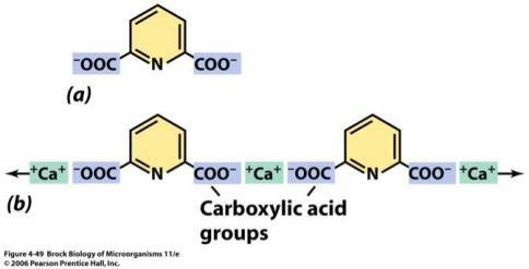
One of the unique compounds found in endospores is dipicolinic acid. Dipicolinic acid is an organic compound that is commonly found in bacterial endospores (about 5 to 15% dry weight of endospores). The dipicolinic acid then forms a complex with calcium ions. This complex of dipicolinic acid and calcium is estimated to make up or can make up 10% of the dry weight of the endospore.
The function of the dipicolinic acid-calcium is to bind and collect water for the purposes of the bacterial endospore.
Another function of dipicolinic acid is to slip between the bases that make up DNA and also support DNA to withstand high temperatures.
Small acid-soluble spore proteins (SASPs)

The core of the bacterial endospore contains many proteins called small acid-soluble spore proteins (SASPs). Small acid-soluble spore proteins, abbreviated as SASPs, are proteins that are only produced when cells are sporulated.
The function of these SASPs is to protect DNA molecules from damage caused by radiation, drought and high temperatures. SASPs cause the structure of the DNA molecule (B-DNA) to become more compact (A-DNA) so that it does not undergo mutations when exposed to UV and is also not denatured when exposed to high temperatures.
Endospore Formation Process
The process of forming endospores is called sporulation. This sporulation usually begins when the cell enters the stationary phase. These cells change either morphologically or physiologically, especially in preparing themselves for the formation of endospores. Some types or types of bacteria are also capable of autolysis of vegetative cells, while for some Other types of bacteria are not able to do this, so the endospores remain in the cell vegetative. The formation of bacterial spores naturally is not well known. However, we can or can trigger bacteria to form spores. Heating at a temperature of 60-65°C for 10 minutes or even more is able to trigger the formation of spores. Other factors that are able to trigger the formation of bacterial spores are the provision of reducing agents, low pH treatments, low temperatures, and other chemical agents.
Mechanism of Sporulation
- In the first stage the bacteria form axial filaments.
- The formation of the axial filament did not last long.
- Formation of an asymmetrical septum, it will produce stem cells as well as pre-spore cell candidates. Each cell will receive daughter DNA.
- After that, the pre-spore cell phagocytosis occurs by the mother cell, so that the pre-spore cell becomes a formation called a protoplast.
- The third stage is the development of the protoplast which is called the early spore development (forespore). In the early spore development, peptidoglycan has not yet been formed, so the initial spore form is irregular (amorphous).
- Formation of the cortex (peptidoglycan). The early spores synthesize peptidoglycan so these early spores have a definite shape.
- The formation of peptidoglycan by the initial spore is also known as cortex formation.
- Formation of the wrapper (coat). Spore-early synthesizes layers of spore envelopes. The spore envelope is synthesized either continuously or intermittently, so that it looks like a thickening of the cortex. Cortical material and spore wrapping are different.
- Spore maturation. Bacterial spores synthesize dipocholineic acid and also carry out calcium uptake. These two components include resistance characteristics and also endospore dormancy.
- The final stage is the release of spores. Stem cell lysis takes place, so that the mature spores come out. No metabolic activity or activity occurs until the spores are ready to germinate. The sporulation process usually takes about 15 hours.
Endospore Painting Method with Malachite Crystals
Generally, the endospores in these bacterial cells are identified by differential staining. The Schaeffer-Fulton method is one type of differential painting. With this method, vegetative cells and endospores will choose different colors until they are easily observed.
The Schaeffer-Fulton method involves malachite green dye as the main dye, while as a mordant or counter stain, red safranin is used. The malachite green dye will be forced to seep into the endospore wall by gradual heating.
The nature of this dye which is easily dissolved in water and weakly bound to the cell wall and endospores will facilitate the destaining process. However, due to the water-resistant nature of the endospore, the malachite green trapped inside the endospore wall cannot be rinsed off and remains green. Only then can endospores and vegetative cells be distinguished by safranin dye which will only color the cell wall because in this process, there is no heating.
The illustration of the endospore painting process using the Green Malachite method is as follows: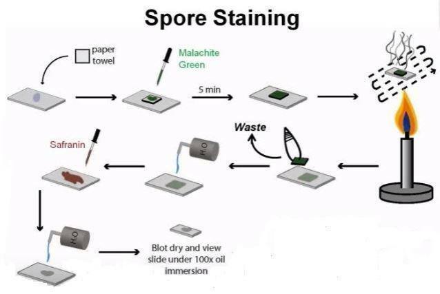
Thus the explanation of the Definition of Endospores, Mechanisms, Processes, Structures, Types and Characteristics, hopefully what is described can be useful for you. thank you
See AlsoDefinition of Saliva Glands (saliva glands)
See AlsoDefinition of Personnel Management
See AlsoEpithelial Tissue