
Definition of Glial Cells, Classification, Types, Structure and Function
Definition of Glial Cells (Neuroglia)
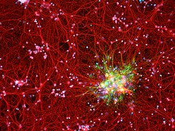
Glia cells (Neuroglia) is a cell that has a function is to support the work of nerve cells. They help in nerve cells so that they can or can carry out their functions properly. These cells can or can be found in the central nervous system or also in the peripheral nervous system. It is estimated that in our brain, the number of glial cells is half of the number of nerve cells (neurons).
Neuroglia is derived from the word 'nerve glue' which was first introduced by Rudolf Virchow in 1854. These neuroglia are composed of all kinds of cells that as a whole support, protect and also play a role, namely: as a source of nutrition for nerve cells (neurons), both in the central nervous system (CNS) or also in the peripheral nervous system (SST). These glial cells play a very important role in supporting neuronal activity. These cells are also very important for the structural integrity of the nervous system as well as for the normal function of neurons.
These neuroglia are support cells for CNS neurons, whereas for Schwann cells they perform this function in the PNS.
Classification of Glial Cells (Neuroglia)
These neuroglial cells, found in the parenchyma of the brain and spinal cord, are broadly classified as:
- Macroglia, these are of ectodermal (nerve) origin, consisting of astrocytes, oligodendrocytes, and glioblasts.
- Microglia, these originate in the mesoderm.
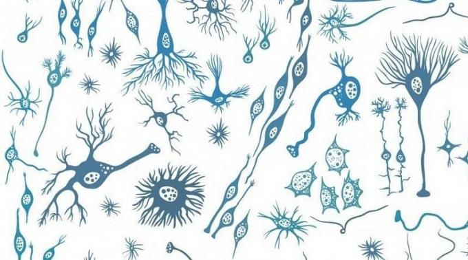
Structure and Types of Glial Cells (Neuroglia)
There are 2 types of glial cells:
1. Glial cells in the central nervous system
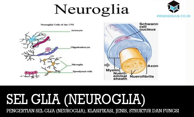
In this central nervous system, there are 4 (four) glial cells including:
1. astrocytes
Astrocytes are so named because they are shaped like stars (astro which means "star", sit is "cell"), is the most abundant glial cell. These cells have important functions, including the following:
- The main “glue” (glia means “glue”) of the CNS, these astrocytes hold neurons together in correct spatial relationships.
- These astrocytes function as scaffolds in guiding these neurons to their final destination during fetal brain development.
- These glial cells trigger the brain's delicate blood vessels to undergo anatomic and functional changes that play a role in the formation of the blood-brain barrier.
- Astrocytes are important in the repair of brain injury and in the formation of nerve scar tissue.
- These cells have a role in the activity of neurotransmitters. Astrocytes absorb and also decompose glutamate and gamma-amino butyric acid (GABA), which are excitatory and inhibitory neurotransmitters, respectively.
- These astrocytes also absorb excess K+ from brain ECF during action or action potential activity the high one beats the ability of the Na+ pump – the K+ returns the K+ that comes out into the neurons. (It should be noted that the K+ leaves the neuron during the descending phase of the action potential.) By absorbing the excess K+, the astrocytes will assist in maintaining the appropriate brain ECF ion concentration for normal nervous excitability.
- Promotes synapse formation and modifies synaptic transmission.
There are two types of astrocytes:
- These protoplasmic astrocytes are abundant in gray matter. These cells have cytoplasmic projections that extend over the entire surface of the cell. Sometimes these protrusions end up in small blood vessels as smaller branches forming "perivascular feet". In the cytoplasm this can or can be shown grains called gliosomes.
- These fibrous astrocytes, on the other hand, are more abundant in the white matter. to see the difference is that with protoplasmic astrocytes it can be seen from the protrusion that is longer and also straight with little branching. Within these projections there is also an image of filaments.
2. Oligodendrocytes
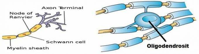
These oligodendroglia are smaller than astrocytes with shorter cytoplasmic branches and fewer branches (oligo = few). The nucleus is small, and the cytoplasm around the nucleus is scanty, appearing as a perinuclear periphery. Golgi complex contains ribosomes, microtubules and neurofilaments.
These cells are mainly located in the gray matter which is closely related to the perikaryon of neurons (they are. cells). perineural satellites) as well as in the lesser amount of white matter lying between the bundles of axon. Others are located close to blood vessels (perivascular).
The function of these oligodendroglia is to form myelin sheaths in the CNS and serve as support cells. The leaflike cytoplasmic branches of the cell bodies extend spirally around the nerve fibers. Each of these oligodendroglia has several branches so that it can or can form myelin sheaths around several adjacent nerve fibers.
These oligodendrocytes form the insulative myelin sheath around the CNS axons. These oligodendrocytes have several elongated projections, each of which wraps (like an omelette) a piece of the axon between neurons to form a myelinated segment.
Oligodendroglia or oligodendrocytes like astrocytes have long cytoplasmic cylinders and are glial cells that are responsible for producing myelin in the CNS. Each oligodendroglia surrounds several neurons and this plasma membrane encloses the protrusions of the neurons to form the myelin sheath. Myelin in PNS is formed by Schwann cells. The function of these oligodendrocytes is to form the myelin sheath in the CNS.
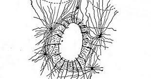
3. Microglia
These microglia are CNS immune defense cells. These “cleansing” cells are the “cousins” of monocytes, a type of white blood cell that leaves the blood and also form the first line of defense in various tissues throughout the body. These microglia originate from the same bone marrow tissue that produces monocytes. During embryonic development, they migrate to the CNS, where these cells remain i.e. until activated by infection or injury.
At rest, microglia are "hairy" cells with many long branches radiating outward. Microglia in this resting state are not just surveillance cells. These cells secrete growth factors in low concentrations, such as nerve growth factor, which help neurons and other glial cells survive and grow. If a problem occurs in the CNS, the microglia then pull the branches, round up, and become very mobile, then move towards the problem area to get rid of all foreign objects or residues network. In an active state, these microglia release destructive chemicals to attack their targets.
4. Ependymal cells
These ependymal cells line the inside of the fluid-filled cavities in the CNS. By the time the nervous system develops in the embryo, the neural tube is hollow, the cavities The initial central portion of this tube is preserved and modified to form the ventricles and canals centralized. These ventricles consist of 4 interconnected cavities in the interior of the brain and also continuous with the narrow central canal which forms a tunnel in the middle of the medulla spinalis. The ependymal cells lining the ventricles contribute to the formation of cerebrospinal fluid, a topic we will discuss shortly. These ependymal cells are one of several types of cells that have cilia. The movement of the cilia of these ependymal cells plays a role in draining the cerebrospinal fluid throughout the ventricles.
The function of these ependymal cells is to coat the inside of the brain cavity as well as the spinal cord cerebrospinal fluid, it has a function as stem cells with the potential to form neurons as well as. cells new glia.
2. Glial cells in the peripheral nervous system
Schwann cells
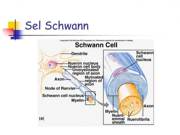
In the axons of the peripheral nervous system, these Schwann cells will allow the occurrence of an electrical signal transduction from the dendrites to the the axon terminal, by coiling the plasma membrane concentrically along the axon known as the sheath. myelin. In the central nervous system, this myelin sheath is formed by oligodendrocytes.
These Schwann cells are unipolar neurons, as in oligodendrocytes, which form myelin and neurolemma in PNS. This neurolemma is a smooth cytoplasmic membrane formed from Schwann cells that enclose the axon fibers of neurons in the PNS, either myelinated or unmyelinated. The neurolemma is a supporting and protective structure for the axon fibers.
Myelin Sheath
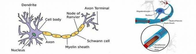
This myelin sheath is a layer that surrounds the axon concentrically and consists of lipids and neurokeratin. In the central nervous system, the myelin sheath is formed by oligodendroglia cells, while in the peripheral nervous system it is formed by Schwann cells.
In the fresh state, this myelin sheath is highly refractile and white (this myelin gives white matter to the white matter of the brain and spinal cord).
Myelin Sheath Formation Process
The process of forming the myelin sheath is initiated by the invagination of the nerve fiber into the cytoplasm of the Schwann cell. The two ends of the cytoplasm of the Schwann cell will then fuse and also wrap around the nerve fiber. This initial fusion site is known as the internal mesaxon. The mesaxons then expand inward to form layers or cytoplasmic lamellae of Schwann cells. The cytoplasm of the Schwann cell then disappears and on both sides of the cytoplasmic membrane it will fuse and also thicken to form a period line. The extracellular membranes of the cytoplasm of the Schwann cells then approach but do not coalesce and form interperiodic lines. At the end of the myelination process, the cytoplasmic wall of the Schwann cell unites for the second time, which is called the external mesaxon.
At the time of the union of the two sides in the cytoplasmic membrane of the Schwann cell there is a failure in several places so that it will then leaving a small amount of cytoplasm entrapped within the myelin sheath known as the Schmidt fissure or notch Lanternan. The fixation using osmium tetraoxide may or may indicate the presence of a Schmidt Lanterman gap. In the CNS, the myelin sheath formation process is similar to that in the PNS, but in the CNS a single oligodendroglial cell can or can make myelin sheaths for several fibers nerve.
This hypothesis regarding the formation of myelin lamellae is also known as the "Jelly Roll" theory.
Myelin Sheath Function
The function of this myelin sheath is like an insulator in an electric wire. The electric current jumps from one node of Ranvier to the next node of Ranvier very quickly (saltatory conduction). Thus the speed of propagation of electrical nerves in myelinated nerves is much faster when compared to unmyelinated nerve fibers.
Functions of Glial Cells (Neuroglia)
The functions of glial cells (neuroglia) include the following:
- Forms the myelin sheath of nerve cells
- Provides Nutrients for Nerve Cells (neurons)
- Maintain body balance
- Participates in the transmission of nervous system signals
Thus the explanation regarding the Definition of Glial Cells (Neuroglia), Classification, Types, Structure and Function, hopefully what is described can be useful for you. thank you
See AlsoDefinition of Negotiation, Purpose of Benefits and Examples
See AlsoDefinition of myelin sheath
See AlsoEditorial Text Material