
The Parts of the Ear with Pictures and Their Functions
Parts of the Ear – Summary of material papers on the ear. On previous occasions we have also discussed about movement system in humans well for now complete material about the parts of the ear.
Hearing is one of the abilities possessed by the human ear that supports communication with each other. In addition, the ear also serves to maintain the balance of the body.
If your ears are disturbed, of course the activities you do are also experiencing obstacles. To find out more, consider the following reviews of the parts of the ear.
Table of contents :
Understanding the Anatomy of the Human Ear:
The human ear consists of three parts, namely the outer ear ( outer ear ), middle ear ( middle ear ), and finally the inner ear ( inner ear ).
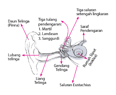
See examples of parts of the ear based on the following three parts:

Sound belongs to the type of longitudinal wave. In order for a sound to be heard, an intermediary medium is needed as a place to propagate, for example, air, water, and others.
In addition, a sound source can be heard by humans under normal circumstances if it has a sound power of 20 Hz to 20,000 Hz. audiosonic sound.
Sounds that are less than 20 Hz are called infrasound and can only be heard by crickets, spiders, and others.
While the sound is greater than 20,000 Hz is called ultrasonic sound and can only be heard by bats and dolphins.
Parts of the Ear and their Functions:
The ear consists of several parts so that it can work properly. The parts of the ear include the auricle, ear canal, ear canal, eardrum, 3 kinds auditory ossicles, 3 kinds of semicircular canals, auditory nerve, eustachian canal, and cochlea cochlea ).
All of these parts have their respective roles so that they can produce information to the brain and finally be able to hear sounds well.
outer ear (outer ear)
This part of the ear is formed from the auricle ( earlobe ) and the external auditory canal ( ear canal or ear canal ). The auricle is formed of elastic cartilage that is firmly attached to the oblique skin.
It serves to capture sound and localize the sound. The part of the auricle forms a depression called the concha and the periphery is called the helix.
The parts of the earlobe consist of:
- helix
- Spiral
- Antihelix
- scaphoid fossa
- triangular fossa
- Antihelical Crura
- Antitragus
- Lobules
- Tragus
Ear canal ( ear canal ) is formed from cartilage and temporal bone. It measures about 4 cm from the tragus to the tympanic membrane (Fig. tympanic membrane ) which is also known as the eardrum and curves in an S shape.
This arch is useful to prevent foreign bodies from reaching the tympanic membrane. There are also mandibular condyles at the front of the ear canal and mastoid air cells at the ends.
There are several sensory nerves in the outer ear, such as:
- Auricular nerve.
- Occipital nerve.
- Ariculotemporal nerve.
- The auricular branch of the pharyngeal nerve ( Arnold's nerves ).
middle ear (middle ear)
The function of the ear in this section is to transmit the sound that has been collected by the auricle to the inner ear.
This part of the ear extends from the cavity to the tympanic membrane to the oval window consisting of the malleus, incus, stapes and many other intricate walls.
For example, lateral wall, medial wall, tagmental wall, and jugular wall. The tympanic membrane is a thin and semi-transparent membrane that separates the outer ear from the middle ear and consists of the pars flaccida and pars tensa.
The manubrium malleus is firmly attached to the tympanic membrane in a concave shape called the. umbo. The higher part of the umbo is called the flaccida pars and the rest is called pars tensa.
There are 3 kinds of sensory nerves in the tympanic membrane, namely the auriculotemporal nerve, Arnold's nerve, and the tympanic nerve branch. On the inner surface of the tympanic membrane is a movable chain of bone called the ossicles.
Namely malleus (hammer), incus (anvil), stapes (stirrup). These bone elements function to transmit and amplify sound waves up to 10 times stronger than air to the perilymph of the inner ear.
In addition, there is also an eustachian tube that connects the middle ear with the upper part of the esophagus and nose ( nasopharynx ). Its function is to equalize the air pressure with the open and close movement.
Important muscles in the middle ear include the stapedius muscle and the tensor tympani tendon. The horizontal section of the facial nerve crosses the tympanic cavity.
Therefore, if there is paralysis of the facial nerves or muscles, it will cause obstructed voice acuity and damage to the inner ear.
inner ear (inner ear)
Inner Air is called the labyrinth cavity which serves to help balance and channel sound to the central nervous system.
This cavity is formed from the osseous labyrinth, which is a series of temporal bones and a membranous labyrinth (membranous sacs and canals). The membranous labyrinth also has a cochlear, vestibular, and semicircular (semicircular) component.
Cochlea ( cochlea ) is an important organ in the inner ear which is shaped like a snail shell. The shape is like a tube bent backwards as far as 2.5 circles with a cone shape at the end.
In this section has 3 kinds of chambers, namely the scala vertibuli, cochlear duct, and scala tympani. In this cochlea, there is an organ of Corti that functions to convert sound waves into nerve impulses.
The vestibule is a link between the cochlea and the semicircular canals. It consists of the saccule and utricle, which are hair cells that maintain the balance of the head against the force of gravity when the body is at rest.
While semicircular is a semicircular channel of 3 different types of canals, namely horizontal semicircular canal, upper vertical semicircular canal, and posterior vertical semicircular canal contains ampulla.
Which serves to determine the awareness of the position of the head when there is rotation or twisting motion.
That's all of the explanation about this ear material, hopefully it's useful...
Also Read:
- Pharyngeal Function
- Mutualism Symbiosis