
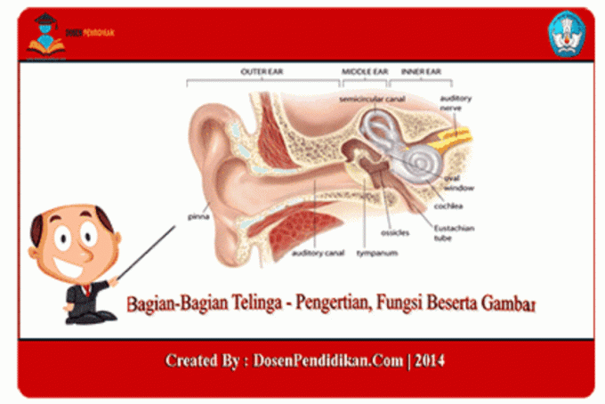
3 Parts of the Ear
For this discussion, we will review: Ear which in this case includes parts, functions along with explanations and pictures, to better understand and comprehend, see the review below.
Definition of Ear
The ear is an organ that is able to detect or recognize sound, and also plays a role in balance and body position in humans. Sound is a form of energy that moves through air, water or other objects, in waves.
Although it is the ear that detects sound, the recognition and interpretation functions are carried out in the brain and central nervous system. Sound stimuli are conveyed to the brain through the nerve that connects the ear and brain, the Vestibulocochlear Nerve.
Also read articles that may be related: 10 Definitions and Anatomy of the Human Brain
Ear Function
The following are several functions of the ear, including:
- Ears as a balance regulator. There is a special structure in the ear organ which functions to regulate and maintain body balance. This organ is connected to the VIIIth brain nerve which functions to maintain balance and to hear.
- Ears as the sense of hearing. The ear can function as a sense of hearing when there are sound waves enters through the outer ear and is received by the brain through the process of hearing which we will explain under.
Also read articles that may be related: Five Senses – Definition, 6 Types, Parts and Functions
Parts of the Human Ear
The human ear consists of 3 parts, namely:
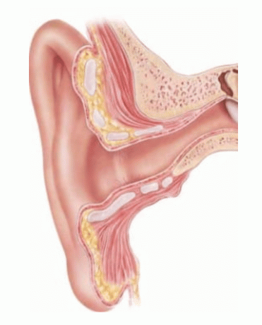
1. Outer Ear
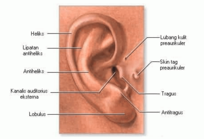
The outer ear consists of:
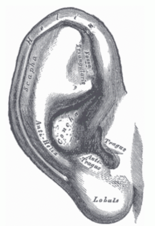
Auricle (Pinna) or Ear Leaf
The pinna or auricula, namely the cartilage leaves which capture sound waves and transmit them to the auditory canal external (neatus), a narrow passage 2.5 cm long that stretches from the auricle to the spinal column tympanic.
The auricle has a skeleton of cartilage covered by skin. In the anterior part of the auricle, the skin is tightly attached to the perichondrium while in the posterior part the skin is loosely attached. The parts of the auricle that do not have cartilage are called lobules.
External Auditory Meatus (MAE) or Ear Canal
MAE is a channel that leads to the middle ear and ends at the tympanic membrane. MAE has a diameter of 0.5 cm and a length of 2.5-3 cm. MAE is a channel that is not straight, but turns from the postero-superior direction on the outside to the antero-inferior direction. Apart from that, there is a narrowing in the medial part called the isthmus.
The lateral third of the MAE is formed by cartilage which is a continuation of the auricular cartilage and is called pars cartilagenus. This part is elastic and covered with skin which is tightly attached to the perichondrium. The skin in this part contains subcutaneous tissue, hair follicles, fat glands (glandula ceruminosa).
Also read articles that may be related: Skin – Function, Anatomy, Structure, Layers, Glands and Their Arrangement
The medial two-thirds of the MAE wall is formed by bone and is called the pars osseus. The skin covering this part is very thin and adheres tightly to the periosteum. In this section there are no hair follicles or glands. Thus it is understandable that cerumen and furuncles can only be found in the lateral third of the MAE.
In the ear area you can find various sensory nerves which are branches of the N.X (N. Arnold), N. V (N. Auriculotemporalis), N. VII, N. IX and branches of N. cervicalis and cervicalis 3 ( N. auricle magnus). Lymph flow from the MAE and auricles goes to the lymph nodes in the parotid, retro-auricular, infra-auricular and glands in the cervical region.
- Ear Canal Skin
The ear canal actually has the same skin layer as the skin layers in other parts of the body, namely it is lined with squamous epithelium. The skin of the ear canal is a continuation of the skin of the earlobe and extends inwards to become the outer layer of the tympanic membrane.
The skin layer of the external ear canal is thicker on the cartilage side than on the bone side. In the ear canal, the cartilage is 0.5-1 mm thick, consisting of the epidermis layer with its papillae, dermis and subcutaneous tissue attached to the perichondrium.
The skin layer of the bony part of the ear canal is thinner, approximately 0.2 mm thick, does not contain papillae, is closely attached to the periosteum without a layer. subcutaneous, continues to be the outer layer of the tympanic membrane and covers the suture between the tympanic bone and the squama bone. This skin does not contain glands and hair. The epidermis of the cartilage part of the ear canal usually consists of 4 layers, namely basal cells, squamous cells, granular cells and horny layers.
- Hair follicles
There are many hair follicles in the outer 1/3 of the ear canal but they are short and scattered irregularly and not so much in the cartilaginous 2/3 of the ear canal. In the bony ear canal, the hairs are fine and sometimes there are glands on the posterior and superior walls.
The outer wall of the hair follicle is formed by invagination of the epidermis which thins when it reaches the base of the polycle, the inner wall of the follicle is the hair itself. The potential space formed is called the follicular canal. There are many sebaceous glands or fat glands in the ear canal and almost all of them lead to hair follicles.
- Sebaceous and apocrine glands
The sebaceous glands in the ear are well developed in the turbinate area, their diameter is 0.5 – 2.2 mm. These glands are often found in the cartilaginous part of the outer ear canal, where they are connected to the hair. In the cartilaginous outer part of the ear canal, the sebaceous glands become smaller, reduced in number and less common completely absent from the skin of the bony ear canal. The sebaceous glands are located in groups in the superficial part skin.
Generally, several adjacent alveoli open into short excretory ducts. These ducts are lined with stratified stratified epithelium which is continuous with the outer covering of the hair roots and with the basal layer of the epidermis, the secretory part of the sebaceous glands in the form of round alveoli with a diameter of 0.5–2.0 mmm. towards the center of the alveoli, a small portion of the cells become horny but increase in size, become polyhydral and gradually fill with fat granules.
Also read articles that may be related: Explanation of the Function of Fat and Its Types
Gradually the core shrivels and disappears, and the cells break apart into fat fragments mixed with the horny sides. This mixture is an oily secretion from the glands, then excreted in the follicular canals and out onto the surface of the skin. Apocrine glands are mainly located in the superior and inferior walls of the ear canal. These glands are located in the middle and lower third of the skin and range in size from 0.5 to 2.0mm. Like sebaceous glands, apocrine glands are formed locally from the outer covering of the hair follicle root.
These glands can be divided into 3 parts, namely the secretory part, the secretory duct in the skin and the terminal duct or epidermal duct component. The circular part of the canal is a tubular structure which rarely branches and consists of an inner epithelial layer, a myoepithelial layer in the middle and a proria membrane on the outside. Surrounding the tabular is dense connective tissue. The epithelium is a single layer varying from cylindrical to very flat cuboidal (flat).
In the cytoplasm, it is usually located supranuclearly and appears as lipoid granules and pigments in varying sizes. The myoepithelium layer is one layer thick, flat cells and contains smooth muscle, forming a continuous covering. gan around the circular part of the gland, and when it contracts it will press on the lumen of the tubule so that secretions will flow go out.
When it reaches the surface of the epidermis, some of these secretions enter the hair follicles and some to the surface free of the ear canal, it will slowly dry out and become semi-solid and become more colored dark. The secretory duct is relatively long and curved and has varying diameters, clearly demarcated from the secretory part of the gland.
Tympanic Membrane

The tympanic membrane separates the tympanic cavity from the external acoustic meatus. The shape is like a cone with an oval base and a conical apex concave medially. The edge of the tympanic membrane is called the margo tympani. The tympanic membrane is attached at an angle by attaching to a groove in the bone called the tympanic sulcus via connective tissue.
Also read articles that may be related: Connective Tissue
The crescent-shaped upper part of the tympanic membrane is called the pars flaccida or shrapnelli membrane. The pars flaccida is thinner and more flexible. The lower part is oval in shape with a pearly white color which is called the pars tensa. The pars tensa is the largest part of the tympanic membrane consisting of a layer of skin epithelium which is a continuation of the acoustic meatus epithelium external, the middle layer (lamina propria) which consists of connective tissue layers arranged circularly and radially and the inner layer which is formed by the cavity mucosa tympanic. The pars flaccida only consists of the outer layer and the inner layer without the lamina propria.
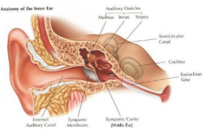
2. Middle Ear
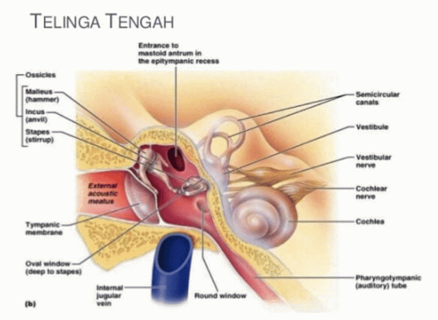
The middle ear is a small space in the temporal bone, separated by the tympanic membrane from the outer ear, the walls of which are further formed by the lateral walls of the inner ear. The cavity is surrounded by a mucous membrane and contains air that enters the pharynx through the auditory canal. This makes the air pressure on both sides of the tympanic membrane the same.
The middle ear consists of three thin bones, called ossicles, which transmit vibrations to the tympanic membrane through the inner ear. The tympanic membrane is thin and semi-transparent and is attached to the malleus, the first ossicle, firmly attached to the inner surface. The incus articulates with the malleus and stapes, the base of the ossicle, which attaches to the fenestra vestibule and points toward the inside of the ear. The posterior wall of the middle ear is irregularly open.
The middle ear consists of:
a. Tympanic Cavum
The tympanic cavity is the most important part of the medial cavity, considering the many structures it contains, namely bones, muscles, ligaments, nerves and blood vessels. The tympanic cavity can be imagined as a box with six walls and its walls border the important organs. The anterior to posterior distance is 15 mm, the superior to inferior distance is 15 mm and the lateral to medial distance is 6 mm, where the narrowest part is only 2 mm.
The tympanic cavity is divided into 3 parts, namely the epitympanum, mesotympanum and hypotympanum. In the tympanic cavity there are:
1) Ossicles consisting of:
- The malleus with its parts, namely the head, column, processus brevis, processus longus, and manubrium malei. The malei head fills the epitympanum, while the other part fills the mesotympanum. The malleus is a hammer-shaped outer bone with a handle attached to the tympanic membrane, while the head protrudes into the tympanic space.
- The incus consists of the head, processus brevis and processus longus. Most of the incus fills the epitympanum and only part of the processus longus fills the mesotympanum. The incus or anvil bone, the outer side articulates with the malleus, while the inner side articulates with the inner side of a small bone, namely the stapes.
- The stapes or stirrup bone consists of the head, column, anterior crus, posterior crus and base. The stapes is attached to the incus at its smaller end, while its elliptical base is attached to the membrane that covers the fenestra vestibuli or oblong window.
The three auditory ossicles are connected to each other by a joint, so that they form a series called the ossicle chain. The base of the stapes closes the foramen ovale through connective tissue called the annular ligament. The ossicle chain and movement of the base of the stapes are very important for the conduction system in hearing function.
b. Muscles consist of M. Tensor tympani which has the function of stretching the tympanic membrane and M. The stapedius has the function of regulating stapes movement.
c. Ligaments, have the function of maintaining the position of the ossicles in the tympanic cavity.
d. The nerve in the tympanic cavity is N. Chord tympani. This nerve is a branch of the pars verticalis N. VII (N. facialist).
The tympanic cavity, which is likened to a box, has the following boundaries:
- The superior wall is a very thin bone of 1 mm and is the boundary between the tympanic cavity (epitympanum) and the middle cranial fossa (temporal lobe). This causes inflammation in the tympanic cavity to spread into the endocranium.
- The inferior wall, in the form of thin bone, is the barrier between the hypotympanum and the jugular bulb. This situation causes inflammation in the tympanic cavity and can cause thrombophlebilitis.
- The posterior wall, there is the aditus and antrum which connects the tympanic cavity with the mastoid antrum and the vertical part of the N.VII canal.
- Anterior wall, there is A. internal carotid, Eustachian tube opening and M. Tensor tympani.
- The medial wall is the boundary between the tympanic cavity and the labyrinth. In this section there is an important structure, namely the pars horizontal semicircular canal which is part of the labyrinth. The horizontal canal of N.VII and the oval foramen are closed by the base of the stapes. The promontory is shaped like a protrusion towards the tympanic cavity which is the first circle of the cochlea. The foramen rotundum is closed by a membrane called the secundary tympanic membrane, which is the boundary between the tympanic cavity and the scala tympani (part of the labyrinth).
- The lateral wall, consists of 2 parts, namely the pars osseus which is the lateral wall of the epitympanum, forms a small part of the lateral wall of the tympanic cavity and pars membranaseus which is also called the membrane tympanic.
b. Eustachian tube
The Eustachian (auditory) tube connects the middle ear to the pharynx. Tubes that are normally closed can open when yawning, swallowing or chewing. This channel functions to balance the air pressure on both sides of the tympanic membrane.
The Eustachian tube is a tube that connects the tympanic cavity to the nasopharynx, has a trumpet shape, 37 mm long. The eustachian tube from the tympanic cavity to the nasopharynx is located in an infero-antero-medial position So there is a difference in height between the mouth of the tympanic cavity and the mouth of the nasopharynx of around 15 mmm.
Also read articles that may be related: Explanation of the Function of the Mouth and Human Oral Cavity
In babies, the Eustachian tube is located more horizontally, is shorter and has a wider lumen, making it easier for middle ear inflammation to occur. The opening in the tympanic cavity is always open, while the opening in the nasopharynx is always closed and only opens when there is an M contraction. Levator and M. The tensor veli palatine occurs when yawning or swallowing.
The function of the Eustachian tube, among other things, is to maintain the pressure in the tympanic cavity the same as the outside air pressure (1 atm) and to ensure ventilation of the air in the tympanic cavity.
c. Mastoid
In relation to middle ear disease, there are 2 important things that need to be learned about the mastoid, namely:
- Mastoid topography
The anterior wall of the mastoid is the posterior wall of the tympanic cavity and external acoustic meatus. The mastoid antrum from the tympanic cavity is connected via the aditus ad antrum.
The upper wall of the mastoid antrum, called the tegmen antri, is a thin wall like the tegmen tympani and is the boundary between the mastoid and the middle cranial fossa. The posterior and medial walls are thin bony walls dividing the mastoid and the sigmoid sinus.
This situation causes inflammation in the mastoid to spread to the endocranium and to the sigmoid sinus so that it can cause inflammation in the brain or thrombophlebilitis.
- Mastoid pneumatization
The process of pneumatization of the mastoid within the mastoid process occurs after the baby is born. Based on growth and shape, 4 types of pneumatization are known, namely
- Infantile, cells that occur as a result of the pneumatization process are very few in number. As a result, the cortex in the mastoid process becomes very thick so that if an abscess occurs it is easier to expand towards the endocranium.
- Normally, the cells that occur expand to such an extent that they almost cover the entire mastoid process. As a result, the cortex in the mastoid process becomes very thin and the abscess easily breaks out, resulting in a retroauricular fistula.
- Hyperpneumatization of the cells that occurs is not only limited to the mastoid process, but also extends to the oszygomatic and even to the pyramidal apex. As a result, inflammation of the mastoid can spread to cause preauricular abscesses and even supraauricular abscesses.
Sclerotic, shaped like infantile type pneumatization. This type of sclerosis occurs due to chronic inflammation in the tympanic cavity and mastoid cavity (chronic otitis media and mastoiditis). As a result, the inflammation spreads more easily towards the ante tegmen, enters the middle cranial fossa and meningitis or brain abscess occurs.
3. Inner Ear
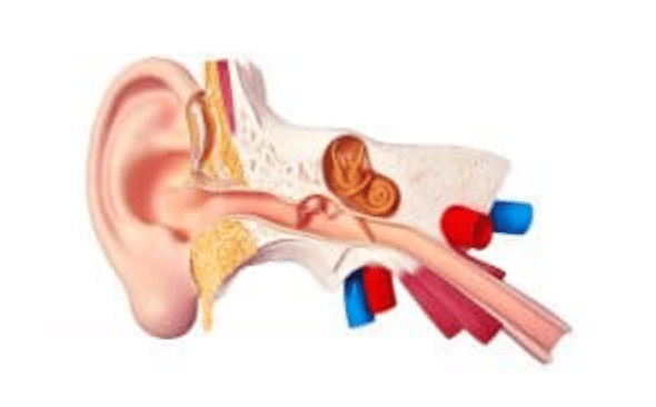
The inner ear is in the petrosal part of the temporal bone which is composed of 2 parts, namely the prominent labyrinth bone and the labyrinth membrane, which is then divided into 3 parts that is:
1. Vestibule
This section is adjacent to the middle ear which passes through 2 holes, namely the vestibuli fenestra which is occupied by the base of the stapes and the cochlear fenestra which is filled with fibrous tissue. At the back there is an estuary that leads to the semicircular canal and at the front there is an estuary that leads to the cochlea.
2. Cochlea
This part is the part of the ear that is important for hearing function. The cochlea is a spiral-shaped canal that forms 2/3 of a turn around the center of the bone which is called the modiolus.
3. Semicircular Canals
Apart from the hearing part, in the inner ear there is the sense of balance control or the vestibular organ. This part is structurally located behind the labyrinth which forms the utricle and saccule structures, as well as 3 semicircular canals or coiled or semicircular canals. The fifth function is to regulate body balance and has hair cells which are naturally connected to the balance part of the auditory nerve.
That's the discussion about Parts of the Ear – Definition, Function and Pictures Hopefully this review can increase your insight and knowledge, thank you very much for visiting.